| |
Ancient DNA (aDNA) analysis
Ancient DNA (aDNA) analysis is a new
research tool with many applications in
fields ranging from genetics through
emerging diseases to forensic medicine. aDNA
techniques provide a unique opportunity to
retrieve genetic information about past
populations unavailable by any other
approach.
The long-term goal of the present research
is to establish a "stratigraphy of genetic
profiles" of the diverse populations which
inhabited Israel in the past, untangling
their inter-group relationships as well as
their genetic links to other cultures in the
Middle East and Europe.
Projects:
a) population origins and movements in the
Southern Levant;
b) origin and spread of genetic disorders in
past populations of the Mediterranean Basin;
Infectious diseases in the past
In ancient Europe, tuberculosis was one of
the most widely prevalent infectious
diseases. Incidence of bone pathology in
skeletal remains from medieval Lithuania
suggests that 18-25% of the population
suffered from the disease. We have detected
the presence of Mycobacterium tuberculosis
in skeletal remains from Lithuania, dated to
the 15th to 17th centuries by amplifying a
part of a repetitive insertion element-like
sequence (IS 6110). DNA of the bacillus has
been identified both in pathological and
normal tissues (bones and even teeth) of the
same individuals, proving hematogenous
spread of bacilli. Moreover, presence of M.
tuberculosis DNA has been demonstrated also
in skeletal remains of individuals without
specific lesions. The results indicate that
a much higher percentage of individuals were
infected than previously thought. Our
findings open the possibility of examining
the actual prevalence of tuberculosis in
ancient populations from collections in
which individuals are represented by single
bones or teeth.
Forensic and archaeological applications
Gender determination of skeletal remains has
considerable importance on forensic and
archaeological studies. However, the
accuracy of identification from morphometric
analyses is limited in the case of
fragmentary remains of adults, or even
complete remains of children. Blind studies
carried out on specimens of known gender,
have demonstrated that while correct
identification may reach as high as 90-95%
for complete adult skeletons, it may fall to
60% or less when dealing with fragmentary
remains or infant skeletons. New
developments in molecular biology have
provided alternative and reliable methods
for gender determination based on
identification of DNA sequences specific to
the X and/or Y chromosomes.

Gender and Burial customs
Today archaeologists are paying increasing
attention to examining social structure
within past societies. Archaeological
studies of gender differences especially in
regard to children have been traditionally
explored through identification of grave
goods considered indicative of female or
male roles. Physical anthropology expands
the study of mortuary practices through
direct identification of sex, even when
grave goods are absent. The reliability of
such analyses varies with the condition of
the bones. For a complete adult skeleton it
approximates 90-95%. However, in fragmentary
remains, or those of infants, the
reliability of sex identification falls as
low as 60%.
Recent developments in molecular biology in
analyzing DNA recovered from ancient bones
have provided reliable methods for gender
determination based on amplification of DNA
sequences specific to the X and/or Y
chromosome (Faerman et
al., 1995). Population structure,
male and female status in past societies,
and gender differences in burial practices
can now be attempted even on fragmentary
skeletal remains as well as those of infants
and children. In Israel, these infant
remains may have been treated with great
care, as for example, the jar burials with
grave goods found at Tel-Teo (Kahila
Bar-Gal & Smith 2001), or
alternately, with complete disregard, like
the infants thrown into sewers in Late Roman
Ashkelon (Smith &
Kahila, 1992).
Out of the 17 burials found at Tel Teo, the
Huleh valley, 10 were infants, with 6 of
them dated to the pottery Neolithic and the
remainder dated to the Chalcholithic and
Early Bronze Age. Nine of these were
subjected to DNA-based sex identification (Smith
et al, 1999). The results were
consistent with all five specimens (of
nine), which yielded amplifiable DNA, being
male.
This method was applied to clarify the
social basis of infanticide in Late Roman -
Early Byzantine Periods (Faerman
et al, 1997; 1998). Skeletal remains
of some 100 neonates were discovered in a
sewer, beneath a Roman bathhouse, which
might have also served as a brothel. Written
sources indicate that in ancient Roman
society infanticide was commonly practiced,
and that females were preferentially
discarded. DNA-based sex identification of
the 43 infant left femurs provided results
in 19 specimens (14 males and 5 females),
indicating that both male and female infants
were victims of infanticide in Ashkelon.
These findings suggested that the infants
might have been offspring of courtesans,
serving in the bathhouse, supporting its use
as a brothel. Later this research was
expanded to Roman Britain (Mays
& Faerman 2001).
Genetic history of modern Israeli
populations
We examined the genetic relationship among
three Jewish communities, the Ashkenazi,
Sephardic and Kurdish Jews, who were
geographically separated from each other for
many centuries. By comparison with Y
chromosome haplotypes of other Middle
Eastern populations we asked how the Y
chromosomes of Jews fit into the genetic
landscape of the region. A sample of 526 Y
chromosomes representing six Middle Eastern
populations (Ashkenazi, Sephardic and
Kurdish Jews from Israel, Moslem Kurds,
Moslem Arabs from Israel and the Palestinian
Authority Area, and Bedouins from the Negev)
was analyzed for 13 binary polymorphisms and
six microsatellite loci. The investigation
of the genetic relationship among three
Jewish communities revealed that Kurdish and
Sephardic Jews were indistinguishable from
each other, while both differed slightly,
yet significantly, from Ashkenazi Jews. The
differences in Ashkenazim may reflect
divergence due to genetic drift during
isolation and/or low-level gene flow from
European populations. Admixture between
Kurdish Jews and their former Moslem host
population in Kurdistan appeared to be
negligible. In comparison with data
available from other relevant populations in
the region, Jews were found to be more
closely related to groups from the north of
the Fertile Crescent (Kurds, Turks and
Armenians) than to their Arab neighbors. The
two haplogroups Eu 9 and Eu 10 constitute a
major part of the Middle Eastern Y
chromosome pool. Our data suggest that Eu 9
originated in Turkey and Eu 10 in the
southern part of the Fertile Crescent.
Genetic dating yielded estimates for the
expansion of these haplogroups that mark the
Neolithic/Chalcolithic Period in the region.
Palestinian Arabs and Bedouins differed from
the other Middle Eastern populations studied
here mainly in specific high-frequency Eu 10
haplotypes not found in non-Arab groups. We
postulate that these chromosomes were
introduced through migrations from the
Arabian Peninsula over the last two
millennia. This study contributes to the
elucidation of the complex demographic
history that shaped the present-day genetic
landscape in the region.
Nebel, A., Filon, D., Weiss, D., Weale, M.,
Faerman, M., Oppenheim, A., Thomas, MG.
2000. High-resolution Y chromosome
haplotypes of Israeli and Palestinian Arabs
reveal geographic substructure and
substantial overlap with haplotypes of Jews.
Human Genetics 107: 630-641.
Nebel, A., Filon, D., Hohoff, C., Faerman,
M., Brinkmann, B., Oppenheim, A. 2001.
Haplogroup-specific deviation from the
stepwise mutation model at the
microsatellite loci DYS388 and DYS392.
European Journal of Human Genetics 9:22-26.
Nebel, A., Filon, D., Brinkman, B., Majumder,
P., Faerman, M., Oppenheim, A. 2001. The Y
chromosome pool of Jews as part of the
genetic landscape of the Middle East.
American Journal of Human Genetics
69(5):1095-112.
Nebel A, Landau-Tasseron E, Filon D,
Oppenheim A, Faerman M. 2002. Genetic
evidence for the expansion of Arabian tribes
into the Southern Levant and North
Africa. American Journal of Human Genetics
70(6):1594-6.
Nebel, A., Filon, D., Faerman, M., Soodyall,
H., Oppenhei, A. 2004. Y chromosome evidence
for a founder effect in Ashkenazi Jews.
European Journal of Human Genetics (in
press).
Health and Disease Studies in Past
Populations
Investigations of human skeletal remains
allow reconstruction of nutritional status
and general health from birth to death.
Physical anthropologists have developed many
techniques for assessing health conditions
and for analyzing their possible causes.
Methods used include measures of health
during childhood, such as stature, location
and severity of dental enamel defects,
cortical bone thickness and the presence or
absence of growth arrest lines. Differential
diagnosis of trauma or disease affecting the
skeleton include morphological,
roentgenological and histological
techniques, chemical and ancient DNA
analyses.
Hand amputation. A complete skeleton of an
adult male, aged 45 years was discovered in
one of the MB-II shaft tombs excavated in
the village located in the Refaim valley.
The right hand was missing and the right
radius and ulna foreshortened and fused
distally.
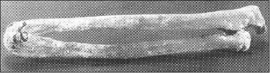 |
| Amputation of the hand. The
distal ends of the radius and ulna
are fused in this 45- year-old male
found in a shaft tomb in Jerusalem
dated to the Middle Bronze I Period
(~3800 B.P.), presumably following
amputation of the hand. Photograph
shows the right radius and ulna with
shortening and rounded fusion of the
distal ends (Bloom et al. 1995). |
No evidence of significant periosteal
reaction, cloacae or distruction was
observed. Radiography demonstrated firm bony
union of the distal shafts of the right
radius and ulna.
 |
| Radiograph showing firm bony
union of the distal ends and absence
of infection. |
The amputation occurred in adult life as
evidenced by the full development and normal
cortical thickness. The appearances are thus
consistent with good healing, following
surgical amputation of a healthy hand or one
traumatically damaged, and is likely to have
occurred at least 1 year prior to death.
This case is the earliest example of limb
amputation originating from Israel (Bloom
et al. 1995).
Skull trephination. During the excavations
at Arad a tomb cave dated to the Early
Bronze age period was discovered which
contained several secondary burials. The
cranial bones of one the individuals, a
young male aged 16-18 years, showed scars
from an old trephination. A large shallow
symmetric depression with shallow sloping
edges was observed on both parietal bones.
 |
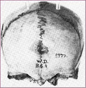
Trephination: Arad, upper view of
the cranial vault. Two symmetric
depressions, resulted from scraping
with a sharp object are seen on the
parietal bones of the skull of a
16-18 male individual from the Early
Bronze Age II period (~4000 B.P.)
(Smith 1990). |
The size and form of the lesion suggest that
this was a trephination carried out by
scraping. The well-healed lesions show that
the death was probably unrelated to the
operation and occurred at least one to two
years post-operation (Smith
1990).
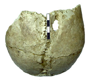
The Goliath Injury. A healed depressed
fracture was found in a Samaritan male aged
about 35 years and dating from the 4th
century B.C. This fracture was located in
the center of the forehead and had a smooth
hemispheric shape.
 |
Frontal view of a cranial vault of a
Samaritan male (2300 B.P.) aged +35
years showing a healed depressed
fracture in the center of the
forehead. It was possibly caused by
a round, smooth missile delivered
with considerable force (Bloom,
Smith 1992). |
A lateral radiograph of this depression
demonstrated it to be a well-healed
depressed fracture of the frontal bone with
no signs of associated osteomyelitis.
 |
Roentgenogram of the fracture. |
The smooth concave appearance of the
depression suggests its causation by a round
smooth missile delivered with considerable
force. This injury may have been the result
of a slingshot wound similar to that of
Goliath who was felled by a stone from the
sling of the young David (Bloom
and Smith 1992).
Ancient DNA analysis is a new research
approach with many applications in fields
ranging from genetics through emerging
diseases to forensic medicine. Our
laboratory has pioneered this field in
Israel. The research has been mainly focused
on the genetics of past and present
populations of Israel, Middle East and
Europe with the main emphasis on population
origins, disease patterns and host-pathogen
relationships.
Anemia. The potential and reliability of DNA
analysis for the identification of human
remains were demonstrated by the study of a
recent bone sample, which represented a
documented case of sickle cell anemia (Faerman
et al. 2000). β-globin
gene sequences obtained from the specimen
revealed homozygosity for the sickle cell
mutation, proving the authenticity of the
retrieved DNA. Further investigation of
mitochondrial and Y chromosome DNA
polymorphic markers indicated that this
sample came from a male of maternal West
African (possibly Yoruban) and paternal
Bantu lineages. The medical record, which
became available after the DNA analyses had
been completed, revealed that it belonged to
a Jamaican black male. These findings are
consistent with this individual being a
descendent of Africans brought to Jamaica
during the trans-Atlantic slave trade.
It is commonly assumed that the endemic
disease load increased with the
establishment of large sedentary
settlements, but there is little direct
evidence of the pathogens involved. Several
hereditary diseases, primarily
hemoglobinopathies, are highly prevalent in
populations residing in regions that have
been infested by malaria.
β-thalassemia is
widespread in sub-tropical malarial regions,
the Mediterranean basin and the Middle East.
It has been suggested that the disease
provided genetic protection against malaria.
Claims for the occurrence of thalassemia in
prehistoric populations have been made on
the basis of skeletal pathology (porotic
hyperostosis). We reported on detecting a
known mutation in the β-globin
gene in the skeletal remains of a child with
severe porotic hyperostosis (Filon
et al., 1995). This study is the
first and so far the only direct proof that
porotic hyperostosis, observed in the
skeletal remains found in the Mediterranean
region, is a result of genetic anemia.
The shift to agriculture and animal
husbandry revolutionized the frequency and
number of contacts amongst people and
between them and animals. It is generally
assumed that, as TB probably occurred as an
endemic disease among animals, the first
human TB cases may have been contracted from
cattle, and Mycobacterium bovis, the agent
of cattle TB, was the most likely first
infecting organism.
Many clinical forms of tuberculosis leave no
specific traces on the human skeleton.
Infectious diseases can be traced through
identification of DNA of a specific pathogen
from human remains. We detected the presence
of Mycobacterium tuberculosis in skeletal
remains dated back to the 1 millennium A.D.
from North-Eastern Europe by amplifying a
part of a repetitive insertion element-like
sequence (IS 6110) (Faerman
et al., 1997). DNA of the bacillus
were identified both in pathological and
normal tissues (bones and teeth) of the same
individuals, and also in skeletal remains of
individuals without specific lesions. The
results suggested that a higher percentage
of the individuals might have been infected
than can be estimated from the incidence of
bone pathology. These findings open the
possibility of examining the actual
prevalence of tuberculosis in ancient
populations from collections in which
individuals are represented by single bones
or teeth.
|
|